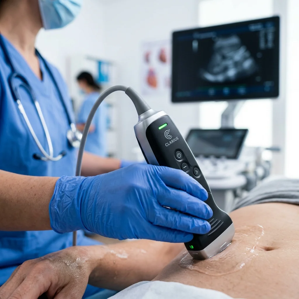
Diagnostic ultrasound stands as one of the most versatile non-invasive imaging technologies in cardiology and pediatrics. To capture high-fidelity readings, clinicians rely on advanced transducer parameters and signal processing setups.
1. Piezoelectric Transducers & Frequencies
Modern ultrasound transducers utilize high-entropy ceramic crystals to generate ultrasonic waves when electrical signals are applied. Selecting frequencies involves balancing penetration depth and resolution:
- • High Frequency (7.5 - 15 MHz): High-resolution superficial imaging, perfect for vascular structures and pediatric cardiac checkups.
- • Low Frequency (2.5 - 5 MHz): Deep acoustic penetration, required for general abdominal checks and adult cardiac diagnostics.
| Clinical Target | Frequency Range | Interoperability Target |
|---|---|---|
| Pediatric Cardiology | 8.0 - 12.0 MHz | DICOM Waveform export |
| Adult Vascular Audit | 5.0 - 7.5 MHz | PACS Database sync |
2. B-Mode Signal Processing
Raw acoustic echo signals undergo filtration, amplification, and envelope detection before translating into pixels on screen. Advanced noise reduction layers isolate tissues from artifact echoes, guaranteeing an accurate diagnostic profile.
3. Color Doppler Imaging
Integrating Color Doppler allows specialists to track moving blood flow structures. Frequency shifts are calculated relative to vascular targets and overlaid as dynamic visual relays on top of standard gray-scale scans.
4. PACS & DICOM Sync
Clinical storage compliance requires saving frames directly to institutional registries. The application exports records via DICOM relays to keep patient imaging histories organized and secure.